There have been lots of buzz around Blake Lively life and career. However, no matter what has been said, it is without a doubt that Blake Lively has gorgeous appearances in the realm of the movie industry. As you know, Blake Lively has been extremely successful in her career in that she has gained lots of awards in his career. However, it has been apparent lately that Blake Lively has been rumored to be no virgin to plastic surgery, one of the most trusted panaceas to cosmetic problems. The rumors about Blake Lively plastic surgery were on everyone’s lips, and they got under the spot light for some time. Perhaps, plastic surgery has been a kind of a must for artists and celebs in as much as this surgical procedure may get them the change and betterment they desire the most. These “betterment” regarding how they appear are so extreme in that plastic surgery gives the impact by changing the facial structure and some other possible parts of their body. As a result, plastic surgery, without great care, can result in disaster. However, Blake Lively assumedly had a really good hand in her plastic surgery. You can see that her appearance is so sparkling. Interested in finding out the truth behind the beauty? Let’s unfold the story behind that fabulous look.
What has the beauty gained through the knife work?
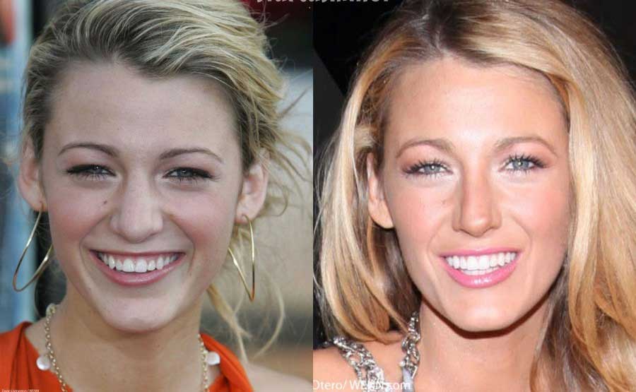
As what commonly happens in the realm of plastic surgery scandals, the rumors about Blake Lively and plastic surgery have been burst by the photos on the internet which posed the difference she has through the knife work. As you can see in the former photos, Blake has rather a bulbous nose with the rather big tip of the nose. However, that look is no longer visible in that you can only spot the slim, pinched, and more pointed nose on her face. Due to that fabulous nose appearance, public assumed that Blake had had the most successful nose job on her face. With all the changes, no wonder that Blake has gained lots of public attention and notice. Also, you may see in the former photo that she has rather tiny breasts on her body. This look, to some great extent, has been escalated through the knife work in that she has had her breasts augmented. Blake Lively has also been rumored to be no virgin to the surgical procedure, particularly breast augmentation. The boob job has sparked a lot of beauty and enchantment on her overall appearance. And now I need you to have long look at her eyelid. You will notice that her eyelid appears so gorgeous as well as sexy. If you compare it with the older photo, there is a slight difference in that her eyelid is as not lifted as that in the newer photo.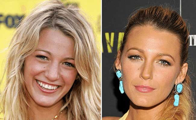
A Queen-like body posed through the plastic surgery
With all the surgeries done very well on her body, Blake Lively has successfully gained lots of public notice and acclaim. The rumors about Blake Lively plastic surgery, no matter whether it is true or not, has made no disastrous impact on her face. Without the surgery, there is no doubt that the actress still looks so gorgeous. With the plethora of her photos on the internet, you will see that her face has been made narrower than before. With all the changes, though quite minor in fact, Blake Lively turns into a massively gorgeous actress. What do you think? Does she look better now or before?
CATEGORY: Beauty
TAG: actress, blake lively, breast, eyelid, nose, plasti curgery
Burton Leon Burt Reynolds has been very well known as one of the most popular American actors and directors. He has starred in a great number of films. Some of his successful movies include Deliverance, Smokey, and The Bandit, as well as White Lightning. With all the frames and sparkling performance shown throughout his career as an actor, no wonder that Burt Reynolds has managed to take a major part in some other movies such as Gator, The Longest Yard, and Boogies Nights. For all the sparkling acts he shown, Burt has managed to scoop some prestigious awards such as Golden Globes Awards for Best Supporting Actor, and The Academy Awards for best actors. Unlike the other celebs and artist who did not gain the same amount of popularity, Reynolds’ life has appeared to be fraught with controversies and scandals. In fact, his rumors have been adorning the public media circus for years. Some of the most public shocking rumors about him were plastic surgery. As you know this particular surgical procedure has gained lots of celebs and socialites’ interest to have undergone the knife work. The rumors regarding Reynolds plastic surgery have been noticed as one of his tremendous speculations. What were the changes he had through the knife work? Well, let’s find them out.
Anything we can notice from his picture?
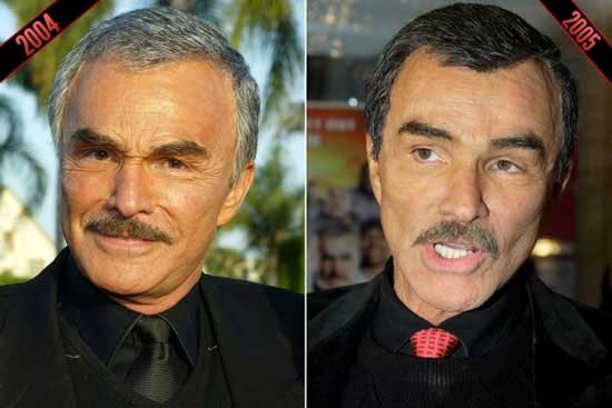
Well, just like what happened to most artists that when first got speculated to have undergone the knife work, the rumors about plastic surgery have been burst by his photos on the internet. Lots of those pictures were labeled horrendously. Let’s try to address the first difference that we can notice. First off, you will see that there are hardly any signs of the most hated enemy in cosmetic, signs of aging. Hardly could you be able to spot any of those signs on Reynolds’ face. His face, not to mention that he has been quite aged, shows no signs of aging. That fountain of youth face has been very extensively speculated to be the outcome of botox injections. This injection has been known by lots of celebs to get them the massively fresh and youthful face. What it exerts on their face is straightened facial muscle which later on will abolish the signs of aging. He has been rumored to have spent a ridiculous amount of money on his knife work. Even if you try to compare the older and the newer photos, there are still no signs of those aging problems. What do you reckon? And there are also some other rumors around his plastic surgery. He was also speculated to have undergone knife work. He was spotted to show some differences on his eyelids. You can see that his eyelid has been made different. Again, public assumed that the plastic surgery was also done on his eyelids.
The Success of Flawless knife work on sparkling face
Perhaps, there will be no ends of having plastic surgery if those having run the surgery felt never satisfied with the outcome of their knife work. With the fascinating outcome he shown on his face, the rumors about his plastic surgery have also been attributed to blepharoplasty. This particular surgery is known to get him extreme change on his eyelid. The other thing we can attribute to his knife work cosmetic escalation is his face fillers. These fillers can be clearly seen on his cheek. Well, what do you say? Even though there are so many pictures about his plastic surgery, it is without a doubt that every one of us has our own judgment on Burt Reynolds plastic surgery.
CATEGORY: Beauty
TAG: actor, burt reynolds, cheek, facelift
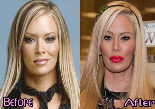
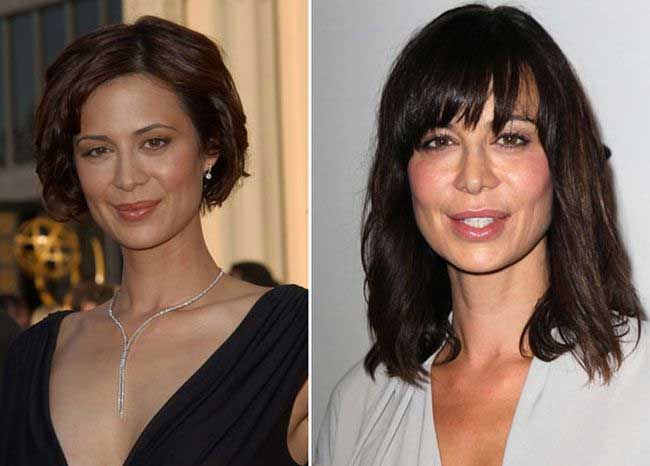
Jenna Jameson was born on April 9, 1974. She is best known as American webcam model, entrepreneur, and former pornographic film actress. She has been called as the world’s most popular adult-entertainment in porn performer. To be exact, she has been popular for her nick name The Queen of Porn. She started her career, the erotic one, in some erotic videos which were released in 1993. She got the career after she had worked as stripped and glamor model in some magazines and tv program. Not until 1996, she had won some other awards on three major adult-movie organizations. No wonder she could procure all those awards in as much as she was indeed truly obsessed with posing her sexy look on camera. In fact, she had won more than thirty adult video awards. In addition, she has been inducted into the X-rater critics organization and adult video news halls of fame. With all the iconic awards that she got throughout her career, no one would be sure that she got all of those awards without any instant and massive effort. Just like what happened to most porn stars, Jenna Jameson was once rumored to have undergone plastic surgery. This surgical procedure has been widely known as the most efficacious and most instant surgical work that can get instant and massive change. The other thing attributed to the knife work is the youthful look it may accrue. Jenna Jameson plastic surgery scandals got under the spotlight for some time, and most people then got really curious if she indeed had the knife work.
The Porn Diva got the best knife work
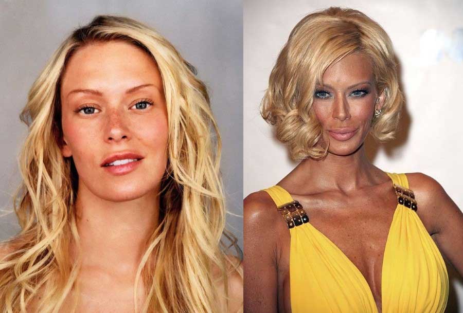
With the career as a porn star, surely Jenna Jameson had lots of demands and pressure to stay sexy and youthful, just like most of her fans desire to see. This paradigm of her career trajectories had caused lots of issues that she was obsessed with the sexy look that she finally decided to have the knife work. The rumors about Jenna Jameson plastic surgery really got public attention. In fact, there were some rumors about her plastic surgeries, claiming that the porn diva had a number of plastic surgeries throughout the year. First off, you can see that, as you can guess, she had breast implants. This surgical procedure has been so popular among female actresses since it can get them a more fabulous look with more massive boob. In the case of Jenna Jameson breast augmentation, you can see that she had a two level escalation in her breast augmentation, which was way too big for her, yet good enough for her iconic symbol as porn diva. This breast augmentation has also been attributed to the look on the entire body. This was because her bodies indicated a massive change in that it got more filled and well built.
The Porn star certainly had nose job as well
Did you think about another surgery she had? Well, of course, she did have another knife work on her face. As what had been posted in Jenna Jameson plastic surgery transformation, you can clearly differentiate her older and newer photos on the internet. Most people would agree that the porn star had a nose job or rhinoplasty. You can see that her nose in the newer photo has appeared much more pointed, pinched, and nicer, she had a smaller and slimmer nose structure, which made her look more fascinating even though this was unnatural.
CATEGORY: Beauty
TAG: breast, jenna jameson, nose job, plastic surgery
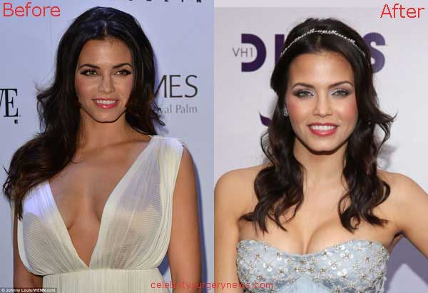
No one would be able to deny the efficacy of the fountain-of-youth surgery which has been widely known to be undergone by artists and actresses in the United States. In the realm of Hollywood movie industry, the rumors about artists taking plastic surgery have been very popular. It is indeed deemed as the most ultimate and instant key to gaining public interest and acclaim. Another thing attributed to the surgical procedure is that it has been known to exert the fountain-of-youth look. Most artists really are dying to stay at the top of their career, and plastic surgery can be the best panacea to their quandary. One of many American artists that were once rumored to have undergone the plastic surgery was Jenna Lee Dewan. Jenna Lee Dewan was born on December 1980. She is best known for her career as American actress and dancer. She has achieved a lot in her career trajectories. She started her career as a backing dancer on Janet Jackson album. Soon after public noticed her talent and passion, which further led her to the other career opportunities when she worked with Missy Elliot, Pink, and Christina Aguilera. Jenna Lee Dewan is also known for her role as Nora in the movie Set Up in 2006. The rumors about Jenna Lee Dewan plastic surgery really came as a whole shock for most people including her fans. The rumors about Jenna Lee Dewan and plastic surgery got under the spotlight and was posted on the media for some time. So, what has, she changed through the rumored plastic surgery? Let’s find them out.

A Real Flawless Knife work?
Most people would agree that Jenna Lee Dewan has a natural beauty on her body. Her face really poses lots of attraction. However, this image has been changed to some great extent due to the existence of her plastic surgery scandals. A lot has been said about the rumors related to her. People were really curious about what she had changed through the surgical procedure. Public curiosity was escalated by the existence of her photos on the internet labeled as Jenna Lee Dewan plastic surgery before and after transformation. First off, you can clearly see that Jenna has, of the most prominent physical change, undergone breast implants. As you can clearly notice in the older photo, Jenna had presumably a B bra cup size. Nevertheless, this size appears to have been escalated in the newer photo in as much as she had bigger boob after the rumors about her plastic surgery got spread. She now is speculated to have a C cup size. You can clearly see that the breast augmentation has been done so nicely. This change sets the sound evidence that the actress indeed had undergone the plastic surgery. Were there any other changes in the rumors about her plastic surgery? You bet!
An Obsession with the cosmetic knife work
After noticing how she has successfully escalated her boob size, Jenna Lee Dewan was rumored to have undergone the other plastic surgeries. Again, Jenna Lee Dewan plastic surgery photos talk a lot in this regard. The other changes that you can easily notice are on her nose and neck. As what has been posted on Jenna Lee Dewan before and after plastic surgery photo, you can see that her nose has been much better structured in the newer photo. This is due to the fact that her nose is much more pointed and more pinched. Also, you may notice that she has a tighter skin around her neck.
CATEGORY: Beauty
TAG: actress, breast, jenna lee dewan, neck, nose, plastic surgery, surgery
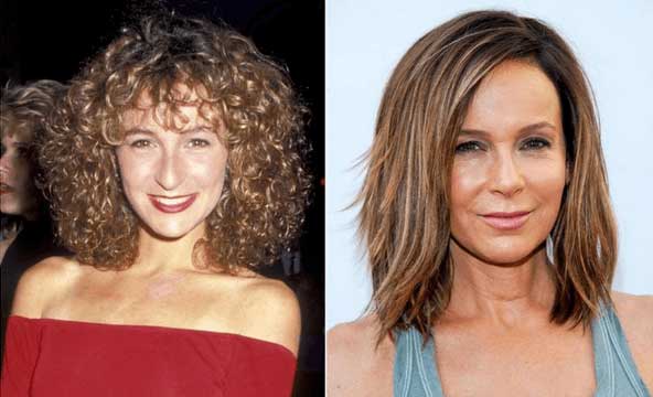
When talking about artists taking plastic surgery, this issue seems to have no end at all. These days, there are more and more rumors being sparked about artist taking plastic surgery to boost their look. In fact, in the realm of movie industry in the realm of Hollywood, there are and have been so many artists, actors, and actresses being rumored to have undergone the knife work to get their look escalated. One of many artists that was once rumored to have been touched by the knife work was Jennifer Grey. Jennifer Grey was born on March 26 1960. She is best known for her career as American actress. In addition, Jennifer Grey is also popular for her role in the movie Ferris Bueller’s Day Off and Dirty Dancing, which were released in the 1980s. For those movie she had played in, Jennifer Grey successfully got nominated for a Golden Globe Award. The other achievement she gained in her career was in 2010 when she won the eleventh season of the American version of Dancing with the Stars. With all the fame and achievement she had gained in her career, people were really shocked by the rumors about Jennifer Grey plastic surgery scandals. Presumably, this was done due to the demand and pressure that she had in her career. However, there were some versions of the issues around her plastic surgery. Some people even thought that she merely had the knife work due to her obsession with looking so young and youthful.
What were the outcomes of the knife work?
Of course, there have been lots of issues about Jennifer Grey plastic surgery transformation. Some people had created so many rumors about her surgery. Perhaps even too many. So let’s explore some of the rumors ever spoken and posted in the media about Jennifer Grey plastic surgery. Of the most prominent change was that the surgery on her eyes. Her eyes apparently had gone through eyelid surgery. This was due to the fact that her eyes look wider and wide open in the newer photo. The other thing which was rumored about her transformation is the nose job. There have been some photos trying to expose the difference on her nose after the surgery. This look is quite different from that in the eyes in the older photo wherein she had rather a bulbous nose with a shorter ridge of the nose. Just like most people assume, the actress managed to get her look escalated through the knife work. Even though she had been rumored to have the surgery, none had ever been said by the actress about the rumors. This was typical reaction most artists would take when facing plastic surgery issues.
Jennifer Grey Surgery Scandals and Rumors
Even though there have been a great number of Jennifer Grey photos on the internet, still the rumors left some rooms for sparking some other allegations about the knife work. As well can clearly see that the actress is supposed to look a little bit old and aged. Most people, commonly, would show some signs of aging at the age of forty or more. However, this does not happen on Jennifer Grey. In the case of her plastic surgery scandals, you can see that her face remains so tight and fresh regardless of the age. There seem to be no signs of wrinkles, sagging, and lines on her face. Nor are there some signs of aging on her face. Due to that fabulous look, which left her on another rumor, Jennifer Grey was then allegedly speculated to have done botox injections. These particular injections have been widely known to get the facial muscle tense and filled.
CATEGORY: Beauty
TAG: botox, eyelid, jennifer gray, nose, plastic surgery, surgery
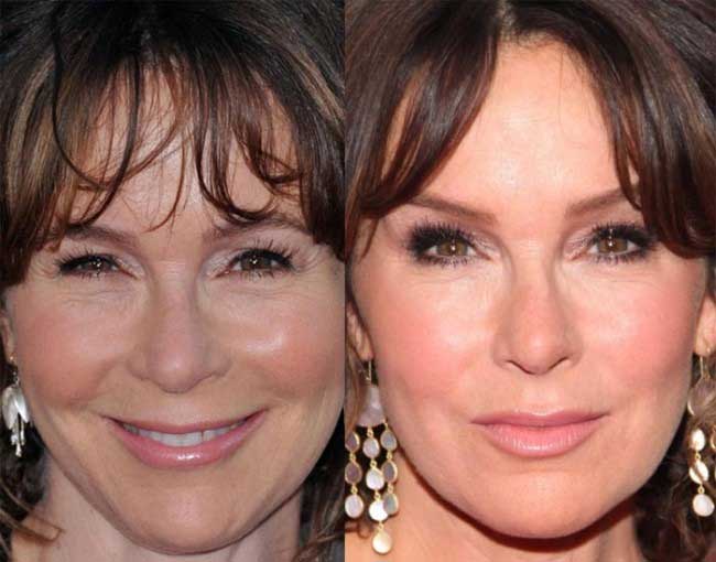
Fabulous Face has been horrendously attributed to the knife work, which is so popular these days, especially in the realm of artists, socialites, and celebs. In fact, this surgery has been well known as one of the most influential keys to gain success and fame. No wonder that there are more and more people who get so fascinated by the knife work and later on decide to undergo the cosmetic surgery. As what is posed in the public media, there are more and more shocking rumors about plastic surgeries. One of the scandals adorning the media circus is Catherina Bell plastic surgery transformation. Catherine Bell was born on August 4, 1968. She is best known as an American actress. Throughout her career, Catherine Bell has taken part in lots of movies such as Death Becomes Her and The Good Witch’s Family. Plastic surgery has been known to exert a very massive change on different parts of one’s appearance. This can be done on forehead, nose, eyelid, lips, cheek, breasts, buttocks; you name it. There is no way that the cosmetic surgery cannot be done on our body. There have been so many versions about Catherine Bell cosmetic surgery. What are they?
source via www.isuwft.com
Did the rumors really unfold the truth behind her beauty?
At first, it is always hard to judge the truth behind a plastic surgery case. However, after years of speculations, the rumors about Catherine Bell and plastic surgery has appeared to be obviously true. This has been caused by the existence of some of her photos on the internet which are meant to pose the difference she has had after undergoing the speculated knife work. This is something which has been common among artist to have themselves exposed to the internet due to the rumored plastic surgery. You can get lots of her photos in which you can easily pose the difference before and after the plastic surgery. First off, you can see clearly that she has had a number of botox injections. Again the desire to have a fountain of youth look has been really prominent in the realm of plastic surgery cases. Catherine Bell was rumored to have not one but several injections on her face. These injections have resulted in her having rather fabulously youthful look. There are hardly noticeable signs of aging such as wrinkles, lines, and sagging. Also, you may think that she has had cheek fillers, which can be true to some extent. In the newer photo, you see that her cheek appears much more filled than that in the former photos. Catherine Bell was rumored to spend a ridiculous amount of money on those surgeries on her face. Were there any other surgeries she had? You bet! The case has yet to suffice here.
The actress surely got way too obsessed with knife work
The plastic surgery apparently also has rather an addictive impact on those who have undergone surgery. This is owing to the fact that whenever one has realized the massively fabulous outcomes that they procure through the surgery, they will be tempted to have more and more knife works done on their body. In the case of her surgery, there are some other rumors about the surgery she had in that she was rumored to have breast implants. Her breast in the recent photo appears much more filled and voluminous than what was posed in the former photo.Also, you may see that she has had minor butt augmentation on her body.
CATEGORY: Beauty
TAG: actress, breasts, buttocks, catherine bell, cheek, eyelid, forehead, lips, nose, plastic surgery, surgery
No one would deny that Catherine Zeta-Jones has a really fascinating beauty posed through her body. In fact, this actress has been very well recognized as one of the most wonderful and sexiest actresses in the United States. Due to the queen-sex-like appearance, Catherine Zeta-Jones has been adorning lots of television program and magazines. Catherina Zeta-Jones was born on September 25, 1969. Even though she is no longer young, she still has the beauty adorning her appearance throughout her life. She has been really popular as an actress. She started her career on the stage when she was really young. After she starred in a number of television films and minor roles in films, such as The Darling Buds of May from 1991 to 1993, she got her prominence in the movie industry with her roles in Hollywood movies which included the phenomenal, The Mask of Zorro in 1998. Also, she has been taking part in some other successful movies such as Entrapment in 1999, Traffic in 200, and some other movies. However, just like what happened to most actresses and actors, there will always rumors and speculations around them. One of the most shocking rumors about her is plastic surgery. With the outstandingly sexy body, she has, no wonder that there are lots of rumors about her plastic surgery. In fact, lots of her rumors regarding the plastic surgery have been one of the most searched news on the internet. Also, you may have noticed that her plastic surgery has been adorning some of the media circuses for some time. However, people, as always, question whether the news is true or not. Let’s find them out.
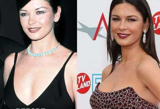
She Certainly got the best knife work
Catherina Zeta is so popular for her, assumedly, naturally sexy look. In fact, the number of her photos on the internet reached massive number. In this case, the rumors about her has been very extremely escalated by the photos labeled Catherina Zeta-Jones plastic surgery. As you can see in the pictures, there are some changes that we can easily attribute to the knife work. First and the most glaring is her breast augmentation. You can see, no matter it is artificial or natural, Catherina Zeta-Jones has a truly fascinating breasts on her body. And, this is one of many things that make her so popular. However, this beauty has to be tainted by her rumors about her taking breast enlargement. Before the plastic surgery, Catherina was speculated to have a C bra cup size, which was rather big already. Nevertheless, that look is no longer visible in her newer photos wherein we are going to see her showing her D bra size. Just like the other breast enlargement, people do think that no matter which one is true, Catherina remains so sparkling with that breasts. The other thing rumored about her plastic surgery is her nose job. Catherina was speculated to have done rhinoplasty. The new nose certainly has escalated her beauty standard even higher. Her nose has appeared much more pointed and pinched.

She had some other surgeries too
There are a great number of rumors about her plastic surgery. You can see in her photos of before and after plastic surgery transformation that she also has botox injections on her face. For someone who has reached forty, it is quite normal that she is about to show some signs of aging, such as wrinkles, sagging, and lines. Whoever, as you can see, Catherina’s face remains so intact and youthful. There are no signs of aging.
CATEGORY: Beauty
TAG: actress, botox, breast, catherine zeta jones, nose, plastic surgery, surgery
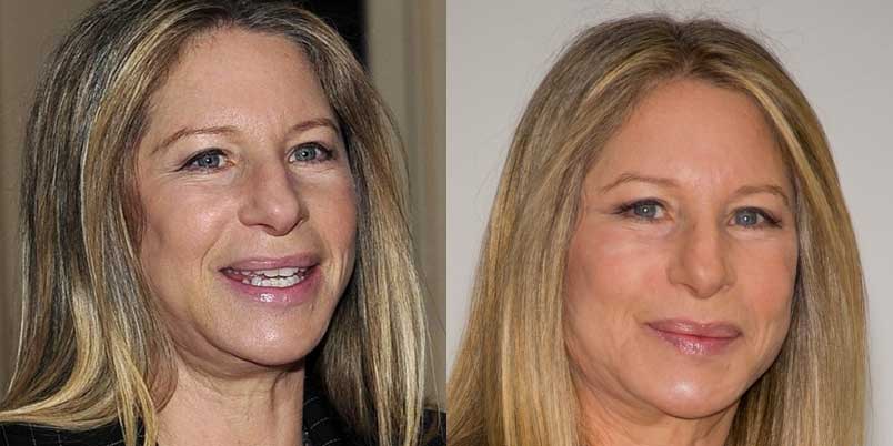
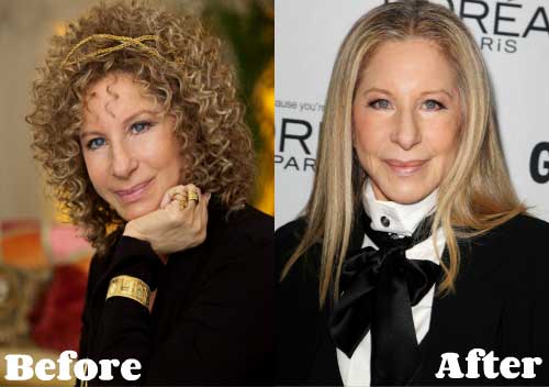
When talking about an artist taking plastic surgery, there seems to be no end of having the end of the very subject. Plastic surgery has been very well known to be the secret panacea to any cosmetic problems. One of many artists that were once rumored to have undergone the plastic surgery is Barbra Streisand. Barbara Joan Streisand was born on April 24, 1942. She has been very popular for her master pieces in the music industry as an American singer, author, song writer, actress, director, and film producer. During her career for more than sixty years, she has turned into a global icon in the music industry. She has certainly achieved a lot in her career in as much as she has been successful to scoop lots of awards throughout her career such as Grammy Awards, Academy Awards, and Emmy Awards. Are those enough? Well, Barbra has also gained some other awards such as Special Tony Award, Day Time Emmy, American Film Institute award, and Kennedy Center Honors. Barbra has been popular for her songs. However, not all the buzz about her is pertinent to the career she has built for years. There have also been some rumors about her. One of the most shocking issues regarding Barbra Streisand was that she had run for plastic surgery. Plastic surgery has been known to be horrendously attributed to those working in the realm of the entertainment industry. And, Barbra Streisand plastic surgery has been tainting her life and adorning the public media circus for some time.
What has been changed through the plastic surgery?
If you are asking about the changes she has had through the knife work, you can easily address the question by looking at some of her photos on the internet. Barbra Streisand before and after plastic surgery photos really have the burst lately. As you can see in her photos in plastic surgery, you will see that Barbra Streisand looks the same throughout the years. For someone who has been over the fifties, that particular look appears so unbelievable yet fabulous. Barbra Streisand appears so youthful and fresh. That miraculous face of Barbra has been rumored to be the result of plastic surgery. To be specific, Barbra’s youthful look has been attributed to be the outcome of botox injections. Apparently, botox injections have been really popular and gained its reliability among artists, celebs, and socialites. You can see that her face shows only very few signs of aging, which commonly include sagging, lines, and wrinkles. The other rumors related to that fountain of youth look is Barbra taking the face lift. Face lift has the potential to abolish any sings of aging by making the facial structure renewed and straightened. These two surgeries are known to be well carried out by the best hand in plastic surgery constellations.
What she said on that rumors
The rumors about plastic surgery have become so tempting to address in the internet and media. And, these speculations may have caused a long rumor about an artist, which is also evident in her plastic surgery. In this regard, Barbra Streisand has been asked, on many occasions, about the plastic surgery attributed to her. And, as most people assume, she mentioned that she had no plastic surgery done on her face. This is quite contrasting to what has been posted in the photos of Barbra Streisand plastic surgery which shows a number of glaring changes. The last thing we can pose in the photos is her nose job.
CATEGORY: Beauty
TAG: actress, barbra streisand, botox, facelift, nose job, plastic surgery, surgery
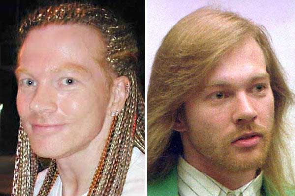
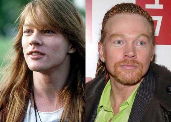
William Bruce Rose Jr, who was raised as William Bruce Bailey, was born on February 6, 1962. He is best known as an American singer and song writer as well as musician. Axl is the lead vocalist and the last remaining member of the hard rock music band named Guns N’ Roses. Because of his extremely powerful and wide vocal pitch as well as energetic performance, Axl has been recognized as one of the greatest rock band singers of all time by lots of media outlets, including Rolling Stone and NME. Born in Lafayette, Indiana, he decided to move to Los Angeles in 1980s. It was in this city where he became even more active in the local hard rock community and took part in a number of bands. In 1985, Axl founded the famous band Guns N’ Roses. It was in this band that he gained the massive success throughout his career. Axl certainly cracked the whole music industry with his action and performance throughout the career. There were myriads of albums the band had created. Even though he has been involved in some rumors and scandals, he somehow kept withstanding all the coming rumors about his life. One of the most shocking rumors about him was regarding plastic surgery. Well, perhaps the paradigm pertinent to entertainment-world demands was also evident in Axl Rose plastic surgery. His rumors about the plastic surgery truly shocked the entire music industry and celebs’ of course. Let’s try to find out the clues behind his plastic surgery scandals.
via evercoream.com
What had the rocker changed through his knife work?
Perhaps this can be rather socking for those who have been well extensively told about the scandals related to plastic surgery. Axl Rose was rumored to have undergone a number of plastic surgeries rather than only one knife work. For someone who has achieved lots of popularity in career, there were so many rumors tainting his life. One of the most searched rumors regarding his plastic surgery was Axl Rose face lift. As the only remaining member of the band, Axl certainly had to be really concerned about how he looked at the stage. This paradigm and then led to the emergence of his face lift scandals. As what has been posed by Axl Rose before and after plastic surgery, you would notice that there were hardly any signs of aging on his face. That smooth and flawless face has unfortunately come as a sign of his having the knife work. Face lift has been very well known to give a massive youthful impact on one’s face, which is apparently nicely done on Axl’s face. The other rumors about his plastic surgery were pertinent to his taking botox injections and face fillers.
No Doubt that he really had the knife work
With the rumors shocking the media circus, the public got more and more amazed by the outcome of Axl Rose plastic surgery. After years of speculations, Axl was caught on some occasions in which he was inquired some questions about his plastic surgery. There were lots of doctors and surgeons who made a claim pertinent to his plastic surgery. However, no matter how many have been said on his rumors on plastic surgery, Axl had made no comments on the very subject, nor had he denied any rumors about the knife work. People then started to fell annoyed with the bunches or rumors regarding Axl Rose plastic surgery. Public was annoyed by the fact that some had burst some excessive comments on the knife work and told them to knock them off.
CATEGORY: Beauty
Barry Manilow is best known as an American singer, producer, and song writer. This singer has been truly popular for his singles such as Mandy, Can’t Smile Without You, and Copacabana. Barry Manilow was born on June 17, 1943. In 1978, there were so shocking albums released by the singer. These singles were on the best seller charts gradually, which was a feat equated by some selects in the music industry such as The Beatles, Herb Alpert, Frank Sinatra, Michael Jackson, Johnny Mathis, and Bruce Springsteen. Barry has recorded a string of Billboard hit singles as well as multi platinum albums. With the buzz of popularity around him, Barry Manilo incredibly turned into a global super star. He has certainly achieved a lot in his career. However, it seems that the popularity he has gained has sparked some rooms for speculations and rumors, one thing which is quite normal for a popular person. Once it was rumored that the singer had undergone knife work to get some changes on his face. Barry Manilow plastic surgery scandals truly came as a total shock in that his rumors were on everyone’s lips at that time. And, as usual, public began to ask the truth behind the rumors regarding the knife work? Anything you can evince on that subject? Let’s find them out.

What has been escalated?
Well, have had any evidence of Barry Manilow plastic surgery? Here under are some of the most obvious ones that we can address by looking at his photos on the internet. When public first heard the news about one’s plastic surgery, they will go on line and search any photos which may pose the difference they can address to the assumed plastic surgery. We can also have the same thing in this regard. Barry Manilow plastic surgery can be well answered by the existence of myriads of his photos. You can see that his face does not change a lot even though he has been quite aged. Hardly will be able to spot any signs of aging. There are only very few sings of aging such as lines, sagging, and wrinkles. People then assume that Barry has had some botox injections on his face. These injections have caused his face to get extremely straightened, which later on demolish any signs of aging. Anything else? You bet..also you may see that he has some face fillers, which are assumedly done on his cheek. These fillers, in conjunction with the botox injections, have caused a very remarkable youthful impact on his face. For someone at that age, what could be better than having a fountain of youth look? Indeed, people argue that the plastic surgery attributed to Barry Manilow is one of the most successful surgery.

What public had thought?
Obviously, there are a great number of perceptions about Barry Manilow and plastic surgery. People assumed that he had the surgery since he wanted to keep his fame intact. This desire has been well realized by counting on plastic surgery. Also, as what has been shown by the photos, you will see that Barry Manilow is so tempted to get some betterment on his face through the surgery. With the constellation of plastic surgery scandals around him, Barry Manilow said that he was so frustrated to get rumored to have undergone the plastic surgery.
CATEGORY: Uncategorized
TAG: barry manilow, botox, cheek, face fillers
Last Posts